
Overview
Achieving a dazzling new year smile prep involves a combination of good oral hygiene, professional dental care, and sometimes cosmetic enhancements to ensure your teeth look their best for the upcoming year.
- Foundational Care: Begin with essential dental check-ups to assess your oral health and address any underlying issues, providing a fresh start for your smile.
- Common Concerns: Learn how to address prevalent smile concerns such as discoloration, alignment issues, or chipped teeth, ensuring a confident appearance.
- Cosmetic Enhancements: Explore various cosmetic treatments available to achieve a truly dazzling smile, from professional whitening to veneers.
- Long-Term Maintenance: Discover effective strategies and habits for maintaining your radiant smile throughout the year, protecting your investment in oral health.
Achieve Your Brightest Smile for the New Year
Preparing for the new year often involves resolutions for personal growth and well-being, and a significant part of that is ensuring your smile reflects your confidence. This section focuses on effective new year smile prep, empowering you to embrace the upcoming year with a radiant and healthy smile. By proactively addressing your oral health, you can unlock a brighter, more confident you.
Investing in your smile is an investment in your overall self-assurance. Whether you’re aiming for a subtle refresh or a more significant transformation, understanding the steps for new year smile prep can make all the difference. From everyday habits to advanced dental procedures, achieving your smile goals can ensure you feel your best as the new year dawns. If you’re considering a procedure like a tooth extraction, reviewing the tooth extraction recovery guide can help ease any concerns.
Essential Dental Check-ups for a Fresh Start
Regular dental check-ups are fundamental to maintaining optimal oral health and are a cornerstone of any new year smile preparation. These appointments allow your dental professional to identify and address potential issues before they become serious problems. Understanding the difference between dental professionals can also be helpful; a dental hygienist vs dentist explains their distinct roles in your care.
Embarking on new year smile prep begins with a foundational step: scheduling essential dental check-ups. These appointments are crucial for identifying and addressing potential oral health issues before they escalate, ensuring your smile is healthy and ready for the year ahead. A thorough examination and professional cleaning provide a clean slate and peace of mind. Learn about the benefits of regular dental checkups to maintain optimal oral health.
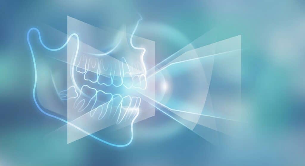
Regular visits allow your dental team to catch problems like early-stage cavities, gum disease, or even signs of oral cancer, which might otherwise go unnoticed. Understanding how often you should visit is also key; the recommended dental cleaning frequency ensures consistent care. Furthermore, dental professionals can assess if new year smile prep requires more than just a standard cleaning, potentially recommending imaging like dental x-rays to get a comprehensive view of your oral health.
Don’t overlook the importance of professional guidance; knowing the top 5 reasons to visit the dentist can motivate proactive care. These check-ups are not just about addressing existing problems but also about prevention, setting the stage for a year of optimal oral health and a confident smile.
Addressing Common Smile Concerns
Preparing your smile for the new year often involves tackling common dental issues that can affect confidence. Addressing concerns like tooth sensitivity or persistent bad breath is a crucial step in your new year smile prep. These issues, while common, can significantly impact your comfort and how you present yourself.
Our team at Heritage Dental Centre can help identify the root cause of these problems and provide effective solutions. Whether it’s a minor adjustment to your daily routine or a more involved treatment, we guide you towards a healthier, more radiant smile. Don’t let common dental concerns hold you back from enjoying your best year yet. Learn more about how to avoid dental problems and maintain optimal oral health.
- Tooth Sensitivity: Experiencing sharp pain when consuming hot, cold, or sweet foods? This often indicates enamel wear or gum recession. Maintaining proper oral hygiene and using specialized toothpaste can help, but professional assessment is key. Learn more about tooth sensitivity causes and relief for more.
- Bad Breath (Halitosis): Persistent bad breath can be embarrassing and is often a sign of underlying issues like gum disease or poor oral hygiene. Regular dental cleanings and consistent brushing and flossing are essential. Discover effective ways to beat bad breath for more.
- Bleeding Gums: While occasional bleeding can occur, persistent bleeding gums are a warning sign of gingivitis or more advanced gum disease. Prompt treatment is vital to prevent further damage and tooth loss. Understand the causes and when to see a dentist for bleeding gums for more.
Understanding Tooth Sensitivity
Tooth sensitivity arises when the protective enamel layer wears away or gums recede, exposing the dentin beneath. This can lead to sharp, sudden pain triggered by temperature changes, sweet foods, or even the air. Consistent, gentle brushing with a soft-bristled toothbrush and a fluoride toothpaste is recommended.
For more persistent sensitivity, professional treatments such as fluoride varnishes or bonding agents can provide relief by strengthening enamel or covering exposed dentin. If you frequently experience discomfort, consult with your dentist to rule out underlying issues like cavities or gum disease, ensuring your common dental issues are properly managed.
Combating Persistent Bad Breath
Bad breath, or halitosis, can be a complex issue with various causes, including food particles, dry mouth, or underlying dental conditions. While brushing and flossing regularly are foundational, sometimes more is needed. Tongue scraping can effectively remove bacteria that contribute to odor.
Staying hydrated is crucial, as saliva helps cleanse the mouth. If bad breath persists despite good hygiene, it may signal more serious problems like gum disease or infections, requiring professional intervention. Exploring options like regular dental cleanings can make a significant difference in maintaining fresh breath throughout the year, and understanding common dental issues and their treatments can empower you to seek the right care.
Cosmetic Treatments for a Dazzling New Year Smile

For those looking to make a significant impact with their new year smile prep, cosmetic dental treatments offer transformative possibilities. These procedures go beyond basic oral hygiene to enhance the aesthetics of your smile, boosting confidence and creating a radiant look for the year ahead. Options range from simple whitening to more comprehensive restorations, all designed to achieve your desired look.
Several advanced treatments can dramatically improve your smile’s appearance. Dental veneers are thin shells custom-made to cover the front surface of teeth, correcting issues like chips, stains, or misalignment. For more significant damage or discoloration, dental crowns can provide a complete aesthetic and functional overhaul. Understanding the various cosmetic treatments for a dazzling new year smile is the first step toward selecting the right option for you. You can learn more about cosmetic dentistry procedures and their benefits.
Exploring these options allows for personalized new year smile prep. A dental restorations comparison: crowns, veneers, implants can help clarify the differences and suitability of each. Consulting with our dental professionals will provide tailored recommendations, ensuring you choose the path that best achieves your aesthetic goals and oral health for the upcoming year. We aim to help you achieve your radiant smile, whether through simple upkeep or advanced cosmetic procedures.
Maintaining Your Radiant Smile Year-Round
Transitioning from new year smile prep to sustained oral health requires a commitment to consistent daily habits. Proactive care at home, coupled with regular professional maintenance, is key to preserving the brightness and health of your smile long after the initial resolutions are made. This ongoing approach ensures your smile remains a source of confidence and well-being throughout the year.
Having worked with individuals aiming for lasting oral health, our team understands that consistent daily habits are the cornerstone of long-term smile success. Beyond the initial boost of new year smile prep, a dedication to fundamental hygiene practices prevents future issues and maintains the aesthetic results achieved. For a comprehensive understanding of what constitutes excellent daily care, explore our oral hygiene guide for more.
Daily Oral Hygiene Routine for Lasting Oral Health
- Brush your teeth thoroughly twice a day for at least two minutes each session, using a fluoride toothpaste. This helps remove plaque and food particles, crucial for preventing decay and gum disease.
- Floss or use interdental cleaners daily to reach areas your toothbrush cannot, effectively clearing debris between teeth and along the gumline. This step is vital in preventing both plaque buildup and its hardened form, tartar.
- Consider using an antimicrobial mouthwash to further reduce bacteria and freshen breath, adding an extra layer of protection for your oral cavity.
- Limit your intake of sugary foods and acidic beverages, as these contribute significantly to enamel erosion and cavities. Opt for water and tooth-friendly snacks when possible.
- Remember to maintain proper brushing techniques, ensuring you cover all surfaces of your teeth without being overly aggressive, which can harm enamel and gums.
Long-Term Smile Preservation Strategies
- Incorporate fluoride treatments or rinses as recommended by your dental professional to strengthen tooth enamel and increase resistance to decay.
- Explore advanced techniques and products that go beyond basic brushing, such as electric toothbrushes or specialized dental aids, to enhance your daily routine. Learn about beyond brushing for a more comprehensive clean.
- Stay informed about the latest advancements in oral care and discuss any concerns or desired improvements with your dentist, ensuring you’re always using the most effective methods for your unique needs.
Your Brightest Smile Awaits This New Year
Embarking on new year smile prep is a fantastic way to boost your confidence and embrace the year ahead with a radiant, healthy smile. By focusing on both routine care and potential enhancements, you set yourself up for success. Remember, a healthy smile is intrinsically linked to your overall well-being, making it a resolution worth prioritizing.
To ensure your smile is truly ready for the year, consider the comprehensive services available to address any concerns. Whether you’re looking to replace missing teeth with durable dental implants in Edmonton or simply seeking advice on maintaining optimal oral health, our team is here to guide you. Understanding the connection between oral health and overall health is the first step toward achieving your smile goals.
Taking the proactive step to enhance your smile for the new year is an investment in yourself. If you’re ready to begin your journey, the best first step is to schedule a consultation with a trusted dental professional. You can find a dentist who aligns with your needs by exploring options for how to choose a dentist that best suits your requirements.